Year 12 Human Biology


Leave a comment

Cystic fibrosis

Cystic fibrosis is a disorder effecting plasma membranes. It is caused by a faulty gene.

One of the proteins involved in active transport across membranes is the CFTR protein, which is made by the CFTR gene. It transports chloride ions out of the cell.

Normal

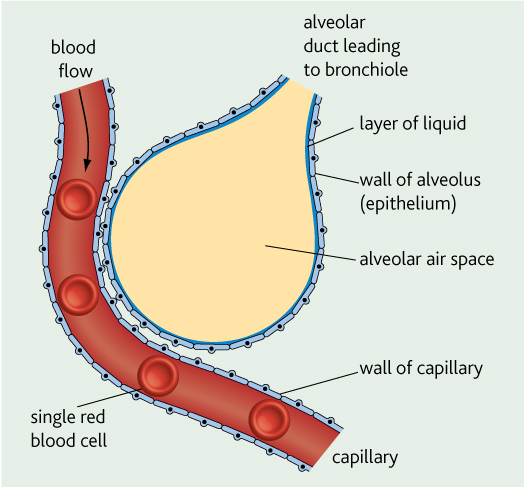
In the respiratory tract, chloride ions are actively transported out of the cells into the mucus which lines the epithelium (remember mucus is produced by the goblet cells). This lowers the water potential of the mucus. This means water moves by osmosis from the cell into the mucus, making the mucus sticky enough to trap dirt and bacteria, but thin enough so it can be moved upwards by the cilia, stopping bacteria settling in the lungs.

Cystic fibrosis

People with CF have a faulty CFTR protein – its tertiary structure is different. Chloride ions cannot be transported out into the mucus. This means the water potential of the mucus increases and water leaves the mucus. This makes it very sticky and thick making it hard for the cilia to move it. This means bacteria settle in the lungs and can cause infections. They develop:

  • coughs and wheezing as they try to force the mucus out.
  • shortness of breath as mucus blocks bronchioles
  • bacteria grow in a layer called a biofilm making it hard for antibiotics and white blood cells to kill them. Lots of phagocytes move to the lungs to destroy them, but end up damaging the lung tissue instead. This lowers the surface area available for gas exchange.
  • thick mucus blocks the pancreatic duct
  • thick mucus blocks the ducts carrying the gametes in the reproductive tracts – can lead to infertility.

Treating cystic fibrosis

  • Chest physiotherapy to help remove mucus from lungs
  • Regular exercise and keeping fit is helpful
  • Antibiotics to help prevent infections
  • Drugs which dilate the airways and thins the mucus
  • Take a high fat, protein and carb diet as they don’t absorb their food properly as well as enzyme capsules to help with digestion due to blocked pancreatic duct.
  • Gene therapy – putting normal copy of CFTR gene into lung cells
  • Lung transplant


2 Comments

Cell structure

Animal cells are eukaryotic – this means they have a nucleus and other membrane-bound organelles (eg. mitochondria, golgi body, endoplasmic reticulum).

Bacteria are prokaryotic – meaning they don’t have a nucleus or any membrane-bound organelles.

You need to know about the following organelles inside a cell:

  • Ribosomes – found in the cytoplasm. This is where enzymes are made that are going to be used in the cell (protein synthesis).
  • Endoplasmic reticulum

The rough endoplasmic reticulum (RER joins up with the nucleus. It has ribosomes on its surface – this is where protein synthesis happens. The RER transports proteins around the cell.

The smooth endoplasmic reticulum (SER) has no ribosomes. Fats are made here.

  • Golgi body – ‘the post office’. Proteins and lipids made in the endoplasmic reticulum are sent to the golgi body. Here they are modified and packaged into vesicles.
  • Vesicles – These carry things out of the cell. They bud off the golgi body and move to the membrane. They then fuse with the membrane and release their contents out of the cell.
  • Mitochondria – supplies ATP (energy storage molecule) which can be used for active transport, muscle contraction as well as other metabolic processes. It has 2 membranes which are folded to form cristae. There is a fluid in the middle called the matrix.

mitochondria

  • Plasma membrane – controls what enters and leaves the cell.

You need to be able to use this to explain how certain molecules are made such as the CFTR protein and mucus.

Mucus is a glycoprotein (a protein attached to a carbohydrate) produced in goblet cells. Proteins made on the RER travel in the RER to the golgi body. Here it gets a carbohydrate attached to it to make mucus. It is put in a vesicle which pinches off from the golgi body and moves to the cell membrane, fusing with it and releasing its contents out of the cell.

cell


Leave a comment

Osmosis

Osmosis – The movement of water molecules from a region of high water potential to a region of lower water potential across a partially permeable membrane.

osmosisWater potential – pure water has a water potential of 0 ψ. This is the highest possible. When other molecules are present in the water the water potential gets more negative. So if the concentration of water molecules decreases (or the concentration of other molecules increases) the water potential gets more negative.

Osmosis happens in animal cells all the time, as our cells are surrounded by fluid.

  • If the solution around the cell has a higher water potential (closer to pure water) than the cell we say the solution is hypotonic. (cell would burst)
  • If the solution around the cell has a lower water potential than the cell we say the solution is hypertonic. (cell would shrink and shrivel)
  • If the solution around the cell has the same water potential as the cell we say the solution is isotonic.


Leave a comment

Diffusion and facilitated diffusion

Diffusion – the net movement of molecules from a region of high concentration to a region of low concentration. This is down a concentration gradient – it is a passive process.

Diffusion often happens across a cell membrane – it happens fastest when there is:

  • a large surface area
  • a short distance
  • a large concentration gradient

Remember, cell membranes are made of phospholipids. This means that small, lipid-soluble molecules pass through easily. The small molecules fit through the membrane. Large and water soluble molecules do not pass through, as the fatty acid tails in the membrane are hydrophobic (don’t like water).


Facilitated diffusion 

facilitated diffusionThis type of diffusion normally uses channel proteins – a type of intrinsic protein. The channels act as a path for molecules to pas through. The channel is filled with water and are specific so only 1 type of molecule can move through it. These allow small water soluble molecules to pass across the membrane

Sometimes carrier proteins are used (see picture to the left). This process does not require energy and molecules move from high concentration to a low concentration – the same as ‘normal’ diffusion.


Leave a comment

The lungs

lung

Each bronchus branches into small bronchioles. At the end of each bronchioles are clusters of air sacs – called alveoli.

Underneath the lungs lies the diaphragm.

Breathing in:

  • Intercostal muscles contract –> ribs move up and out
  • Diaphragm contracts and so flattens

This increases thorax volume and reduces pressure –> air is pulled in.

Breathing out:

  • Intercostal muscles relax –> ribs move down and inwards
  • Diaphragm relaxes and so becomes dome shaped again.

This reduces the thorax volume and increases the pressure –>air is forced out.

Alveoli – where gas exchange happens

alveoli


The inner surface is moist – stops cells drying out. The fluid contains a surfactant which lowers surface tension stopping the alveoli sticking together when breathing in and out. The oxygen also dissolves in this layer before diffusing into the capillaries around the alveoli.

How are alveoli adapted for efficient gas exchange?

  • large surface area – millions of alveoli in the lungs and each one is folded increasing the SA even more.
  • thin permeable surface – epithelium of alveoli is made of very thin flat cells. The wall of the capillary is only one cell thick and the capillary is very close to the alveoli. This all means that oxygen has a very small distance to diffuse across.
  • large concentration gradient – breathing in brings in air with high oxygen concentration and low carbon dioxide concentration. The capillaries are bringing deoxygenated blood to the alveoli containing lots of carbon dioxide. This sets up a large concentration gradient for oxygen to diffuse into the blood and carbon dioxide to diffuse into the alveoli and so we breathe out air high in carbon dioxide and low in oxygen.

How are the lungs kept clean?

The trachea and bronchi are lined with an epithelium (outer layer of cells). This has 2 cells in it:

  • Ciliated cells – have cilia on the edge (hair like structures that are an extension of the cytoplasm). These bend and beat in time with each other and waft any mucus up the trachea, where it can be swallowed.
  • Goblet cells – make and secrete mucus. Mucus is slimy and sticky and traps dust and bacteria that is breathed in.

With the mucus sweeping the mucus away this helps to reduce the chance of respiratory infections.


Leave a comment

Antibiotics

Antibiotics can be used to treat bacterial infections and diseases.

They kill bacteria by interfering with their metabolism.

  • They stop them making new cell walls when they divide, so the cell takes in water by osmosis and bursts.
  • The stop bacteria making proteins. They interfere with their ribosomes and so protein synthesis cannot happen.

Antibiotic resistance

antibiotic resistance


Some bacteria are resistant to lots of antibiotics – these are multi-drug resistant bacteria.

Antibiotic resistance is more likely to develop when antibiotics are widely used. How can this be stopped:

  • Doctors avoid giving antibiotics for minor infections present no danger – if the bacteria are not exposed to the antibiotic resistance is unlikely to develop.
  • Doctors should prescribe antibiotics for viral diseases – antibiotics do not destroy viruses, but if bacteria are present, they might develop resistance.
  • Always finish the course of antibiotics – there may be a few bacteria left after the illness has gone, these will be the bacteria that are most resistant to the antibiotics, so if you don’t finish, they will survive and multiply, passing on the resistant gene and spread to other people.

MRSA is a well known bacteria. It is resistant to the antibiotic methicillin. Hopsitals take care to avoid spreading MRSA in hospitals:

  • contact isolation – wash hands before and after touching patients
  • put patients in a separate room if they have it in their nose so they can’t spread by droplet infection
  • clean surfaces regularly to eliminate it from dust
  • clean beds after a patient has used it
  • put MRSA patients together in a separate room
  • Staff wear clean disposable aprons and gloves, and throw them away after use
  • visitors use alcohol hand rub to sterilise hands before and after visiting


Leave a comment

Tuberculosis

What causes TB?

The bacteria called Mycobacterium tuberculosis. It infects any part of the body, but particularly the lungs as these are easily infected.

It is spread by droplet infection – spread through the air by coughs or sneezes which contain tiny droplets of mucus, containing bacteria. If someone breathes these in, the bacteria enters the lungs. Usually you need to be in close contact for a long time to be in danger.

  • Overcrowding
  • Poor ventilation

These can contribute to the spread of TB.

Meat or milk from infected animals can also spread TB. TB from milk usually infects the bones.

How does it cause disease?

In most people it lies dormant, but can become active years later especially is the persons immune system is weakened (ie. by HIV).

  • Macrophages in the lungs engulf the TB bacteria.
  • The macrophage acts like a shell and keeps the bacteria under control.
  • This means most people don’t develop the disease.

The next stage is when tubercles develop. These are dead lung tissue (fibrous tissue) which surround the bacteria. The bacteria can’t multiply in the tubercles as there is low oxygen and there is a low pH. But they do remain alive. See the picture below of a tubercle.

tubercle

  • The bacteria are surrounded by macrophages,
  • The bacteria that are inside these macrophages are able to divide, this causes the tubercle to get bigger,
  • The bacteria may enter the bronchioles and other parts of the lung as it gets bigger, or enter the blood stream and get to other parts of the body.

The final stages:

  • Inside the tubercles starts to liquefy – the bacteria start to divide very fast,
  • The tissues of the bronchioles die and break open leaving cavities in the lungs,
  • So the bacteria can spread quickly in the lung.

What are the symptoms?

  • Develop a cough – lots of mucus and blood in the airways which blocks them and so need coughing up.
  • Fever and loss of appetite
  • Difficulty in breathing – if alveoli are damaged, there will be  reduced surface area for gas exchange, because of all the fibrous tissue you won’t be able to breathe in as deeply.
  • Skin infections can develop

How is it diagnosed?

  • X-ray – to look for tubercles which show up as shadowy areas.
  • Too prove its TB – a skin test is needed – the Mantoux test. An antigen from Mycobacterium is inserted in the skin, if the red and swelling around the antigen is bigger than 15mmm it is TB, or the person has already been infected with it in the past.

How do we treat TB?

  • A mixture of antibiotics. If just one is given, the bacteria will probably develop antibiotics resistance to it. It is hard for the drug to penetrate the tubercles because of the fibrous tissue so they need to be taken regularly over 6 months. The tubercles act as a reservoir of infection.
  • It can be prevented by a BCG vaccine.

Risk factors of TB

  • People with HIV are more likely to get infected with TB (because their immune system is compromised and so less likely to fight off the infection).
  • Global travel has increased its spread.
  • Drug-resistant forms have developed – hard to treat.
  • More homeless people, living in overcrowded, poorly ventilated accommodation.


Leave a comment

Salmonella

Salmonella is a bacteria that causes food poisoning.

Where does it come from?

Found in intestines of many animals, so may be in animal faeces.

How is it spread?

  • By eating or drinking food contaminated with faeces that has the bacterium in it.
  • Unvaccinated chickens may have it in their eggs.
  • Eating raw food that contains the bacteria.

Large numbers of the bacteria are needed to cause disease, so it is normally caught if food has been left to stand around in warm, damp conditions.

How does it cause food poisoning?

  • The bacteria invades the lining of the small intestine,
  • They rapidly divide here,
  • When they die, they release an endotoxin when they burst open,

The endotoxin causes the symptoms of food poisoning:

  • vomiting
  • diarrhoea
  • abdominal pain
  • fever
  • dehydration

What is the treatment?

They are often left to run their course. Drink lots of water and rest.

In the young or elderly, oral rehydration therapy is given. This is a drink containing sugar and salts, which replaces mineral ions lost in the body.

Antibiotics are only given in severe cases.

How can we prevent food poisoning?

  • If food is frozen, allow it to completely thaw and then cook all the way through – this will kill any bacteria. If it’s cooked before defrosting completely, some parts of the chicken might not heat up enough to kill the bacteria,
  • Wash your hands before handling food,
  • Dispose of bleach old dish clothes regularly,
  • Don’t mix raw meat with anything else.